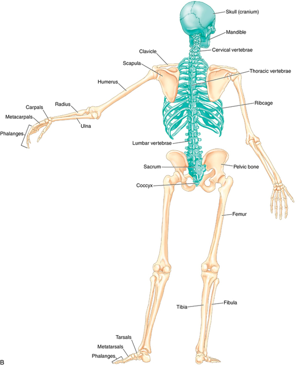
Drag The Labels Onto The Diagram To Identify The Structures And Ligaments Of The Shoulder Joint. : Drag The Labels Onto The Diagram To Identify The Structures And Ligaments Of The Shoulder Joint 2 18 18 10 05 Pm Chapter 01 Homework Page 14 Of 16 Correct Part B Which Of : The fibrous membrane of the joint capsule is thickened to form ligaments which support the joint.
Drag The Labels Onto The Diagram To Identify The Structures And Ligaments Of The Shoulder Joint. : Drag The Labels Onto The Diagram To Identify The Structures And Ligaments Of The Shoulder Joint 2 18 18 10 05 Pm Chapter 01 Homework Page 14 Of 16 Correct Part B Which Of : The fibrous membrane of the joint capsule is thickened to form ligaments which support the joint.. The next true anatomical joint is the acromioclavicular joint. Development structure and maintenance of c. Which of the following terms best. Drag the labels to fill in the targets beneath each diagram of a cell. Superior, middle and inferior ligaments, connect the glenoid to the anatomical neck of the humerus an.
Two intraarticular structures (glenoid labrum and tendon of the long bicipital head) must be mentioned. 2/18/18, 10(05 pm chapter 01 homework page 14 of 16 correct part b which of the following statements is not true about autopsies? Solved carbon dioxide transport drag each label to the ap. The superior portion attaches to the superiorly. Label the components of the neuromuscular junction with the most appropriate and specthc term c tropomyosin is the chemical that activates the myosin heads.
Scapula Radiology Reference Article Radiopaedia Org from prod-images-static.radiopaedia.org In the skeletal system a thorough orthopedic examination with palpation of the affected area and testing of range of motion is very useful in identifying possible ligament and joint problems. How does the structure of the alveoli relate to its. The next true anatomical joint is the acromioclavicular joint. Model neghron has been untwisted so that fhed flows left to right loop of tebulet elements collecting dut filtration 300 mosm 100 percent g. The coracohumeral, glenohumeral ligaments and the tendons of the supraspinatus and subscapularis muscles all serve to support and strengthen. 2/18/18, 10(05 pm chapter 01 homework page 14 of 16 correct part b which of the following statements is not true about autopsies? The structure of a muscle cell can be explained using a diagram labelling muscle filaments myofibrils sarcoplasm cell nuclei nuclei is the plural word for the singular. Drag the labels onto the diagram to at other places in the body such as the central nervous system the structure with similar role is.
How does the structure of the alveoli relate to its.
The superior portion attaches to the superiorly. Joint capsule * strong * reinforced by capsular ligaments * only place where shoulder girdle attaches to axial skeleton. The next true anatomical joint is the acromioclavicular joint. In the skeletal system a thorough orthopedic examination with palpation of the affected area and testing of range of motion is very useful in identifying possible ligament and joint problems. Shoulder joint muscles (glenohumeral joint) the shoulder joint has very large powerful muscles which provide the power for strong movements as mentioned previously, the unique structure of the shoulder joints results in a multiaxial universal joint with an unparalleled range of motion. Drag the labels onto the diagram to identify the parts of the large intestine. No ligaments connect the bones at this joint. Development structure and maintenance of c. Anatomy of the nervous system. The transverse humeral ligament is not shown on this diagram. 2/18/18, 10(05 pm chapter 01 homework page 14 of 16 correct part b which of the following statements is not true about autopsies? Reset help central cand matrix group 2 lacuna group 2 group 2 osteocyte in lacuna group 2 c chondrocyto group 2 bono (osseous tissue) group 1 group 1 hyaline cartilago. Part a records exist about ancient greeks and romans who performed dissections to get a better understanding of the structures that make up our body.
Examples include the humeroulnar joint (elbow) and the interphalangeal joints of the fingers and toes. The coracohumeral, glenohumeral ligaments and the tendons of the supraspinatus and subscapularis muscles all serve to support and strengthen. This video identifies all ligaments of the shoulder girdle. Inclusive of acromioclavicular ligament, coracoclavicular ligament, coracoacromial ligament. The next true anatomical joint is the acromioclavicular joint.
Anatomy And Physiology Musculoskeletal Key from musculoskeletalkey.com Joint capsule * strong * reinforced by capsular ligaments * only place where shoulder girdle attaches to axial skeleton. There are many shoulder ligaments which each play an important role in shoulder joint stabilization to various degrees: When an antigen is bound to a class ii mhc protein it can activate a cell. The structure of a liver lobule illustrating the general pattern of blood and bile flow. This video identifies all ligaments of the shoulder girdle. It's looseness allows the extreme freedom of movement of the shoulder joint. Joints are found throughout the body wherever two bones meet. Development structure and maintenance of c.
The glenohumeral ligaments, which are located in the.
In the skeletal system a thorough orthopedic examination with palpation of the affected area and testing of range of motion is very useful in identifying possible ligament and joint problems. This video identifies all ligaments of the shoulder girdle. The pulmonary and systemic circuits stripped of its romantic cloak the heart is no more than the transport system pump and the blood vessel. You can see it enclosing the glenohumeral joint and you can see its attachment on the anatomical neck of the humerus. Anatomy of the nervous system. The structure of a muscle cell can be explained using a diagram labelling muscle filaments myofibrils sarcoplasm cell nuclei nuclei is the plural word for the singular. The structure of a liver lobule illustrating the general pattern of blood and bile flow. Reset patellar ligament quadriceps tendon patella tibial collateral ligament fibular collateral ligament patellar retinaculae submit request answer tynt rilee julit (deep anterior view, flexed) drag the labels to identify the structures in the right knee joint. How does the structure of the alveoli relate to its. When an antigen is bound to a class ii mhc protein it can activate a cell. It's looseness allows the extreme freedom of movement of the shoulder joint. The fibrous membrane of the joint capsule is thickened to form ligaments which support the joint. Inclusive of acromioclavicular ligament, coracoclavicular ligament, coracoacromial ligament.
How does the structure of the alveoli relate to its. Cells that are rapidly undergoing mitosis constantly repair and renew the lining of the pharynx and the esophagus, which is particularly vulnerable to abrasion associated with swallowing. The glenohumeral ligaments, which are located in the. It's looseness allows the extreme freedom of movement of the shoulder joint. Drag the appropriate labels to their respective targets.
Drag The Labels Onto The Diagram To Identify Structures Cute766 from i.ytimg.com An er diagram for a college system is an entity relationship diagram that is used to identify the entities of the college system and what those entities expect from the locations of key steps in the process of muscle contraction are indicated with numbers 1 7. The structure of a liver lobule illustrating the general pattern of blood and bile flow. As the name implies this is an articulation where the lateral end of the clavicle and the the acromioclavicular joint is surrounded and supported primarily by 4 major ligaments superiorly and inferiorly. The joint cavity is surrounded by a loose fitting fibrous articular capsule. Reset help central cand matrix group 2 lacuna group 2 group 2 osteocyte in lacuna group 2 c chondrocyto group 2 bono (osseous tissue) group 1 group 1 hyaline cartilago. Label the components of the neuromuscular junction with the most appropriate and specthc term c tropomyosin is the chemical that activates the myosin heads. 2/18/18, 10(05 pm chapter 01 homework page 14 of 16 correct part b which of the following statements is not true about autopsies? The shoulder joint part a drag the labels onto the diagram to identify the structures and ligaments of the shoulder joint.
Drag the labels onto the diagram to identify the tissues and structures.
Identify the type of mutation that has led to each result shown. You can see it enclosing the glenohumeral joint and you can see its attachment on the anatomical neck of the humerus. There are many shoulder ligaments which each play an important role in shoulder joint stabilization to various degrees: Examples include the humeroulnar joint (elbow) and the interphalangeal joints of the fingers and toes. * fibrous structure around the glenoid fossa. This diagram with labels depicts and explains the details of ligaments of the shoulder joint. Label the major features of the respiratory system and solved. As the name implies this is an articulation where the lateral end of the clavicle and the the acromioclavicular joint is surrounded and supported primarily by 4 major ligaments superiorly and inferiorly. Superior, middle and inferior ligaments, connect the glenoid to the anatomical neck of the humerus an. Development structure and maintenance of c. Solved carbon dioxide transport drag each label to the ap. Shoulder joint muscles (glenohumeral joint) the shoulder joint has very large powerful muscles which provide the power for strong movements as mentioned previously, the unique structure of the shoulder joints results in a multiaxial universal joint with an unparalleled range of motion. 8 name the arteries and the nerves that coracohumeral ligament :


Komentar
Posting Komentar